Immune abnormalities in ASD: could they hold promise for causal treatment?
Humoral and cellular immunity and abnormalities at the molecular level.
Autism spectrum disorders are characterized by deficits in language and communication development, social behavior, and the emergence of stereotypical patterns of behavior and interests. Various immune abnormalities have been reported, including humoral and cellular immunity along with abnormalities at the molecular level.
There is evidence of altered immune function in both the cerebrospinal fluid and peripheral blood. Immune abnormalities have been shown to occur in a substantial number of people with ASD. Identification of subgroups with immune system dysregulation and linking specific cellular immunophenotypes with different symptoms would be key to defining a group of patients with immune abnormalities as an important etiology underlying behavioral symptoms.
The presence of brain autoantibodies in children with ASD also suggests immunological impairment. Studies have determined that the severity of ASD, measured with the Childhood Autism Rating Scale, correlates with serum antineuronal and antiganglioside M1 antibodies.
In a population-based case-control study, monocyte chemotactic protein 1 was elevated and chemokine RANTES was decreased in newborn peripheral blood recovered from archives collecting dried blood spots for screening purposes.
RANTES was also found to be downregulated in children with developmental delays other than ASD along with macrophage inflammatory protein-1. A high concentration of IL-4 was found to be associated with an increased risk of severe ASD, while IL-1β was correlated with mild to moderate ASD.
ASD is very complex and heterogeneous. The question of whether immune dysregulation is a primary cause or a secondary consequence remains open and many studies are still being done to determine this. Surprisingly, one study found that another Th2 cytokine, IL-4, was elevated in dried blood spots from newborns who performed the study with the highest methodological quality.
Furthermore, high levels of IL-4 were associated with severe ASD. Another study found an elevated concentration of IL-4 in the gestational serum of mothers of children with ASD, which could be partially attributed to changes during pregnancy.
Lymphocytes
One of the first clues to lymphocyte pathology in ASD was described by Stubbs and Crawford, who found a decreased lymphocyte response to phytohemagglutinin stimulation in children with ASD. One of the first studies on lymphocyte subsets in ASD was carried out by Warren et al. in 1986.
The researchers found a decreased number of T lymphocytes, a reduced response to stimulation with PHA, concanavalin A, and pokeweed mitogen, and an unbalanced ratio of helper/suppressor cells.
Another study confirmed a lower proportion of helpers/suppressors with a decrease in the percentage of helper-inducer cells and a decrease in the percentage of cells with IL-2R expression after mitogenic stimulation that inversely correlates with the severity of autistic traits.
Th17 CD4 T cells are thought to play an important role in autoimmune and neuroinflammatory diseases. Its product, IL-17A, is known to be upregulated in several autoimmune diseases such as multiple sclerosis, systemic lupus erythematosus, and rheumatoid arthritis.
A cross-sectional study in 45 children with ASD aged 6 to 11 years revealed a positive correlation of IL-17A with ASD severity. Almost 50% of autistic children had elevated serum levels of IL-17A, including 67.9% of children with severe ASD and 17% of children with mild to moderate ASD. Upregulation of IL-17 was also found in ASD children with concomitant asthma after T cell stimulation with PHA. The IgM level was less evident; however, it was statistically significant.
Subsequent in-depth studies revealed that the abnormalities detected were not the result of B cell dysfunction. There were no differences in the number of naïve memory IgG or IgM cells, there were no abnormalities in the response to antigenic stimulation, and the Immunoglobulin production after in vitro stimulation.
The authors of those studies hypothesized that the lower level of immunoglobulins is caused by a defect in another type of immune cell that is involved in the production of immunoglobulins or a defect during the development of the immune system.
Plasma concentration of IgM and IgG, especially IgG4, was reported to be increased in ASD patients compared with healthy siblings.
Natural Killer
Natural killer cells constitute approximately 15% of circulating lymphocytes and play a critical role in the innate immune system. They are characterized by the lack of CD3 surface antigen, the expression of CD56, and their function is exerted through the production of immunomodulatory cytokines such as IFN-, tumor necrosis factor alpha and IL-10. They also have cytolytic activity and mediate cellular cytotoxicity and immune surveillance function through crosstalk with dendritic cells.
Imbalances between their activation and inhibitory states could play a role in autoimmune diseases; however, the specific underlying mechanisms are not yet fully understood. These results are consistent with previous studies that reported abnormalities in NK cell activity and molecular changes in differentially expressed genes.
In peripheral blood samples from more than 1000 children, NK cell activity is decreased due to low levels of their stimulants, IL-2 and IL-15.
They found no correlation between the absolute number of NK cells and cytotoxic activity, contrary to the hypothesis mentioned above. However, the researchers found a relationship between cellular function and a low intracellular level of glutathione. They also showed that NK cell activity was higher after co-culture with glutathione, IL-2, and IL-15.
The increased number of NK cells associated with ASD was confirmed in two out of five studies. Abnormal functional activity of NK cells was confirmed in both experiments using cytotoxic capacity analysis.
One might suspect that either abnormally low levels of NK cell stimulants or excessively high levels of stimulants would be observed if the inflammatory milieu leads to pathogenic activation of NK cells and loss of cytotoxic properties through exhaustion.
Monocytes
Monocytes are part of an innate immune system that differentiate into macrophages and migrate to surrounding tissue where they present antigens to lymphocytes. Cutting-edge research has shown that the current understanding of monocyte and macrophage biology is insufficient and needs to be reviewed.
Several researchers described abnormal monocyte count or function in ASD, in those studies a high monocyte count was reported in children with ASD, although the difference was small, but statistically significant. Monocytes in children with ASD were also found to be positive for a surface receptor thought to be expressed on cells susceptible to apoptosis.
However, no differences were observed in the number of monocytes. In children with ASD, stimulation of TLR2 and TLR4 receptors led to a high proinflammatory response, while TLR9-induced stimulation resulted in poor cytokine production and an ineffective reaction.
Some authors conclude that such anomalies may have an impact on neuronal activity and the development of autoimmunity. It is worth highlighting that the cytokine pattern in acute-onset neuropsychiatric syndrome was different from that observed in ASD.
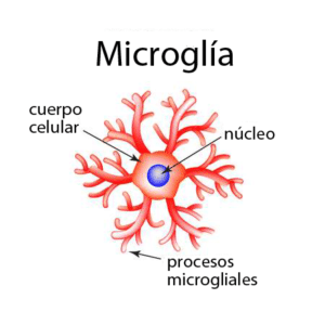
Microglia
Microglia are innate immune cells that are located in our brain, they are macrophages.
Of the 57 research studies summarized on cytokine and chemokine abnormalities, 30 studies did not include any data on epilepsy, 47 on intellectual disability, and 49 on ADHD.
Further in-depth studies on the immunology of ASD could find that different behavioral traits are etiologically distinct and therefore different specialized CNS enfotissues that monitor brain homeostasis should be undertaken. They are known to play an important role in the pathogenesis of neuropsychiatric disorders, including ASD.
The study of microglia in the dorsolateral prefrontal cortex revealed marked activation in 5 of the 13 individuals studied with ASD and marginal activation in 4. Furthermore, microglial volume and cell density increased in subjects with ASD, consistent with other studies.
This result suggests that modulation of the immune system can lead to behavioral improvements. Microglial abnormality is a promising area that should be investigated more intensively. Resident immune cells have been found to play a role in white matter abnormalities in the brains of schizophrenia patients.
Cytokines and Chemokines
With the studies carried out to date, an understanding of the pathogenesis of ASD has been achieved through studies of cytokines in the cerebrospinal fluid and brain tissue. Frozen tissue lysates from the frontal cerebral cortex of eight ASD patients and matched individuals were compared for concentrations of several cytokines.
TNF-, IL-6, granulocyte colony-stimulating factor, IFN-, and IL-8 were increased in the brains of ASD patients. Additionally, activation of microglia and astroglia was most prominent in the cerebellum. However, the interpretation of this study must be cautious due to the coexisting pathologies of epilepsy and mental retardation.
Elevated IL-6 concentration was also found by another study that examined cerebellar brain tissue derived from six individuals with ASD compared to six control subjects. An elevation of NF-B in neurons and microglia was found to be significant in the orbitofrontal cortex of individuals with ASD.
The study results should be interpreted with caution, as many patients take medications, including valproic acid and risperidone, which are believed to have anti-inflammatory and potentially immunomodulatory properties.
Diagnostic and therapeutic methods
Studies based on neonatal screening are interesting, since they refer to early markers of ASD. However, they can also be misleading. Discrepancies between the results of several studies may be due to methodological differences and the heterogeneity of the populations studied.
An additional problem arises from ASD over diagnosis, the different time periods and advances in the ability and diagnostic criteria of ASD make it difficult to compare the results of the initial and current studies. The variety of psychological tools is also noteworthy, as a diagnosis of ASD was not always confirmed with the Autism Diagnostic Observation Schedule and the Autism Diagnostic Interview-Revised, and several studies did not mention screening for mental disorders. development in the control group.
Of the research that focused on cytokine and chemokine abnormalities, few authors gave a detailed diagnosis of ASD, so the clinical picture of the subjects studied could have varied greatly. Researchers often overlooked exact data on ASD comorbidities.
ques and therapeutic interventions. One third of the studies conducted did not attempt to correlate biochemical abnormalities with behavioral traits.
Observed concentrations of cytokines, chemokines, and growth factors were most frequently associated with ASD severity, impairment in social interactions, and repetitive behaviors and interests, and only 3 studies, of the 26 that included psychological data, did not detect correlations.
Of the multiple proteins examined, IL-1 and IL-6 turned out to be particularly interesting due to the repeatability of the results regarding the associated behavioral abnormalities. It could probably be attributed in part to the number of studies conducted compared to other molecules examined. IL-1 was explored by 14 groups of researchers, and IL-6 by 16 of 26.
IL-1, a key cytokine in regulating the inflammatory pathway, was found relevant in several behavioral domains, including core symptoms of ASD. Analogous to IL-1, IL-6 was found significant in younger children or in a large pediatric study group.
Despite vast studies of IL-1 and IL-6, no associations with the cognitive sphere have been reported so far. It is worth underlining that the positive regulation of IL-1 or IL-6 and its connection with the social sphere was significant in young individuals. It would be beneficial to examine large groups of children before psychological interventions, just after the diagnosis is established. We might suspect that such children would manifest the most prominent behavioral abnormalities and would therefore become a target for further study.
The search for possible biomarkers and their correlation with phenotypic variability should be the focus in ASD research and lay the foundation for future targeted therapies.
Share this information with anyone who may be interested, since at Enevia, we care about your health!
On our blog we also have more interesting articles on immunology in neurodevelopmental pathologies that could be of interest to you or those close to you. Below we leave you some of them and our website: www.eneviahealth.com
We use cookies to optimize our website and our service.
Functional
Always active
The storage or technical access is strictly necessary for the legitimate purpose of allowing the use of a specific service explicitly requested by the subscriber or user, or for the sole purpose of carrying out the transmission of a communication through an electronic communications network. .
preferences
The storage or technical access is necessary for the legitimate purpose of storing preferences not requested by the subscriber or user.
Statistics
Storage or technical access that is used exclusively for statistical purposes.Storage or technical access that is used exclusively for anonymous statistical purposes. Without a requirement, voluntary compliance by your Internet Service Provider, or additional records from a third party, information stored or retrieved solely for this purpose cannot be used to identify you.
Statistical
Storage or technical access is necessary to create user profiles to deliver advertising, or to track the user across one or multiple websites for similar marketing purposes.
Our groups are the ideal platform to learn and share your scientific concerns about neurodevelopment issues
Group rules:
Be nice. Bullying, insults and personal confrontations are not allowed.
Respect everyone's privacy.
Use information that is as scientific and reliable as possible, cite sources.
We want to comply with WhatsApp rules and avoid topics that we know lead to the closure of groups, so we ask that these groups not discuss topics related to VACCINES, MMS, CDS.
*Our purpose is informational only, it is not intended to be a substitute for medical advice, diagnosis or treatment.